Syndromes coronariens aigus (ACS)
The nomenclature of anterior infarction can be confusing, with multiple different terms used for the various infarction patterns. The following is a simplified approach to naming the different types of anterior MI. The precordial leads can be classified as follows:
Septal leads = V1-2
Anterior leads = V3-4
Lateral leads = V5-6
The different infarct patterns are named according to the leads with maximal ST elevation:
Septal = V1-2
Anterior = V2-5
Anteroseptal = V1-4
Anterolateral = V3-6, I + aVL
Extensive anterior / anterolateral = V1-6, I + aVL
OMI (STEMI)
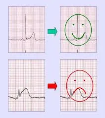
Péricardite (vert) vs OMI (rouge)
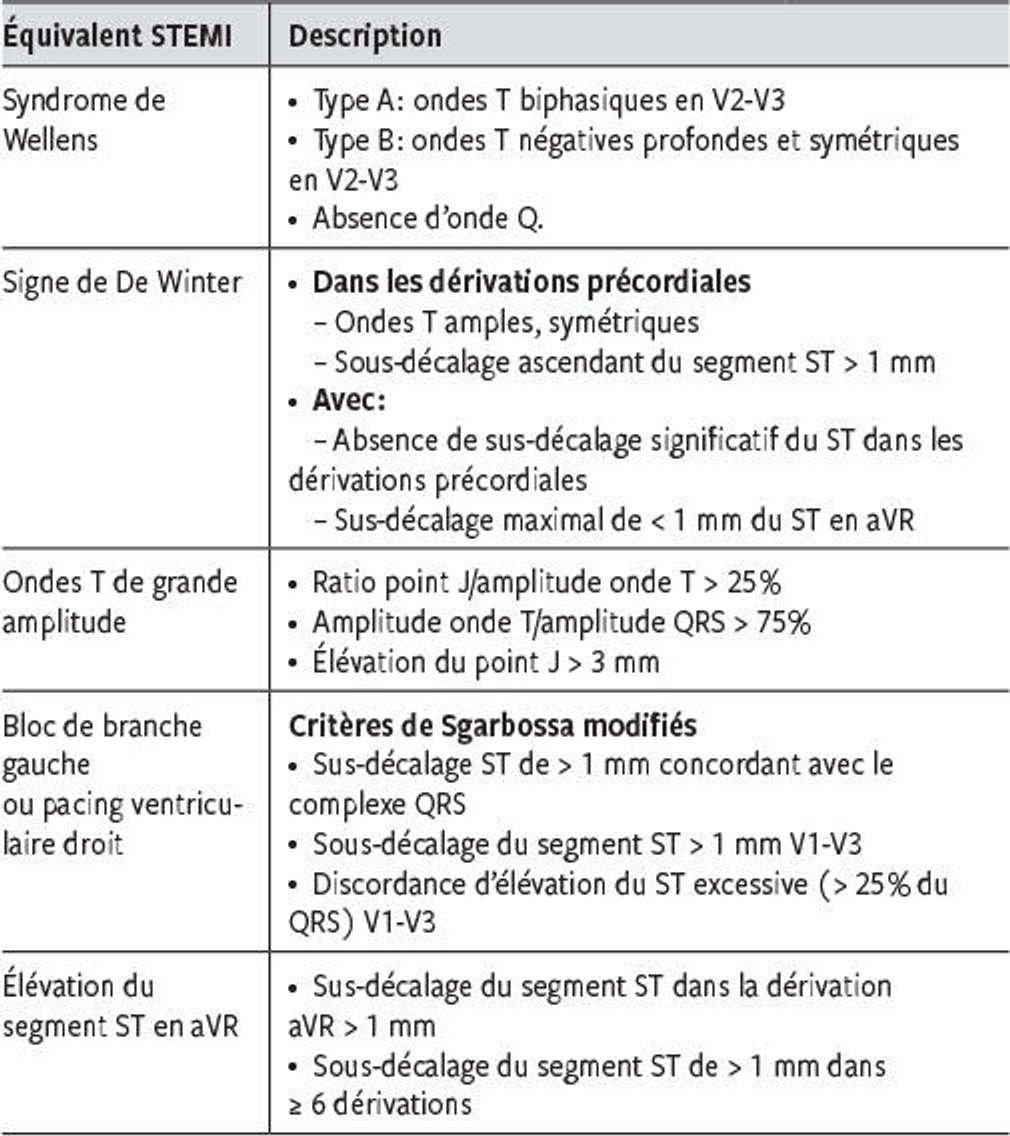
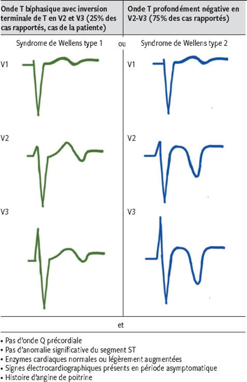
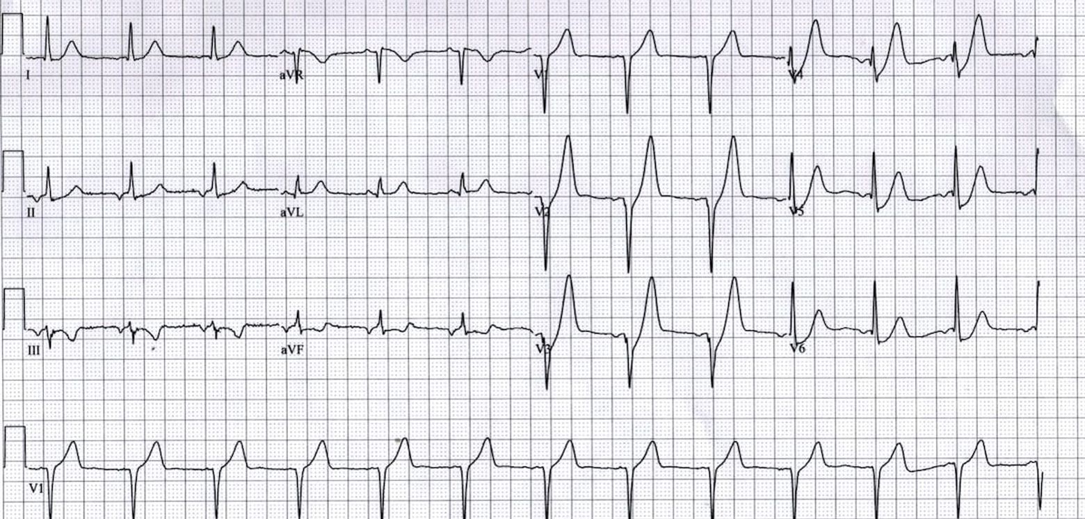
Syndrome de Wellens (syndrome de l’IVA/LAD)
Symptômes résolutifs et anomalies ECG au niveau de l’onde T dans les dérivations (V1)-V2-V3, qui reflètent un stade de préinfarctus avec une obstruction critique de l’artère interventriculaire antérieure (IVA).
Syndrome de Wellens: ondes T biphasiques dans les précordiales
Evolution progressive d’ondes T biphasiques (type A) en ondes T profondément inversées (type B)
Syndrome de Wellens
TTT: cardiologie interventionnelle pour éviter la survenue d’un infarctus antérieur massif
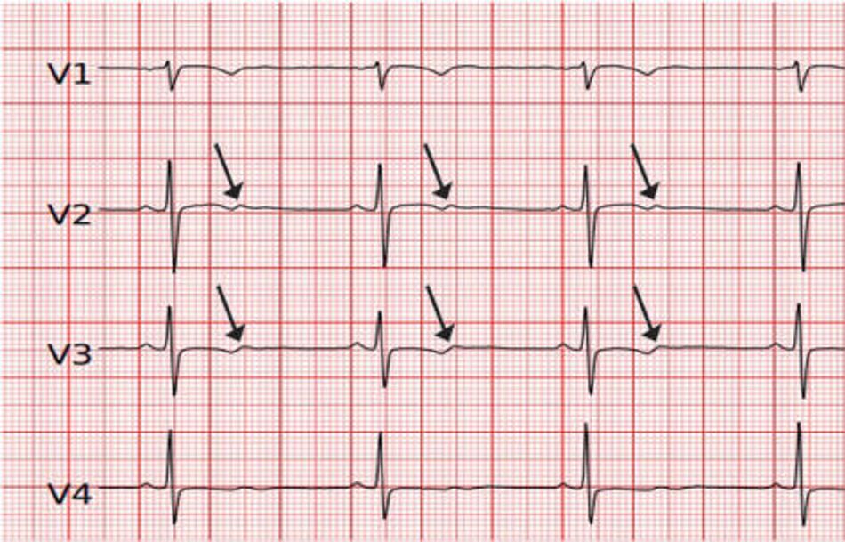
Ondes T de De Winter
Anterior STEMI equivalent that presents without obvious ST segment elevation.
(ST depression) and peaked T waves in the precordial leads
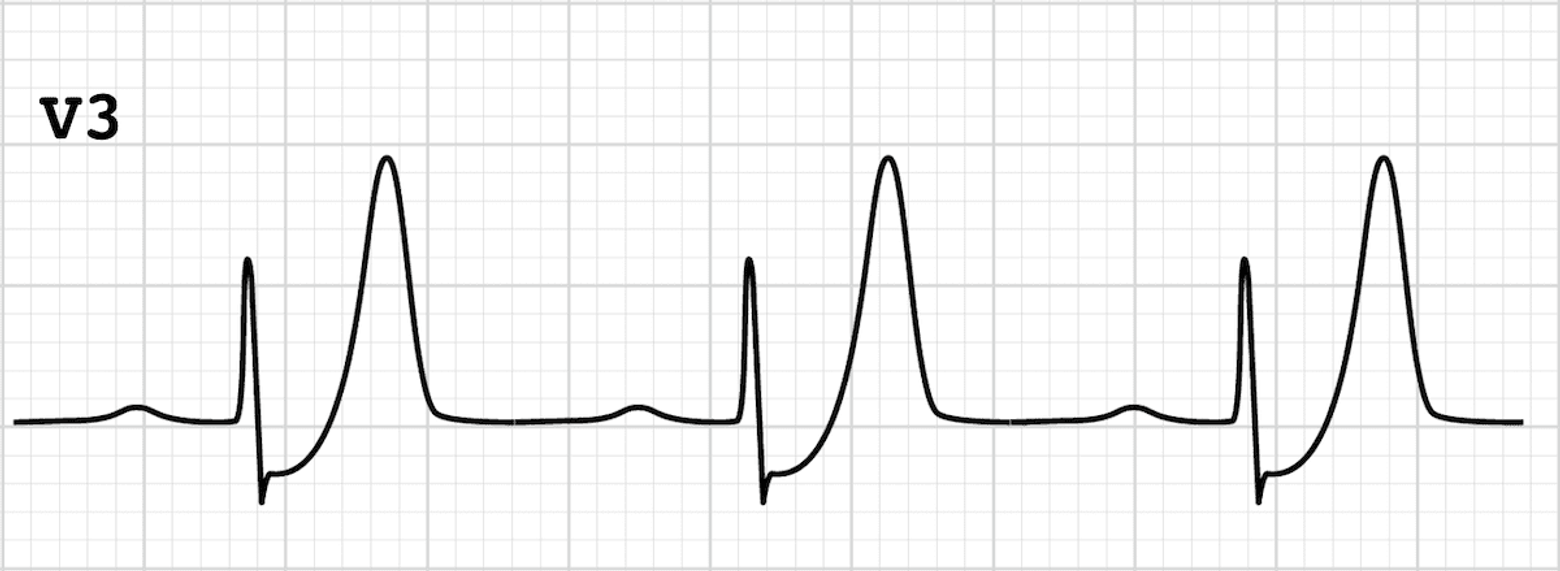
Ondes T de De Winter
Ondes T de De Winter
En général les ondes T de De Winter persistent plusieurs heures ou jusqu’à reperfusion de l’IVA, alors que les ondes T hyperaiguës sont transitoires et évoluent en STEMI classique; la prise en charge reste la même
Although tall symmetrical T waves have been recognized as a transient early feature that changes into overt ST elevation in the precordial leads, in these patients this pattern was static, persisting from the time of first ECG until the preprocedural ECG was performed and angiographic evidence of an occluded LAD was obtained (i.e., 30 to 50 minutes). — https://www.nejm.org/doi/full/10.1056/NEJMc0804737
South african flag sign
High lateral STEMI is associated with a pattern of ST elevation caused by acute occlusion of the first diagonal branch of the left anterior descending coronary artery (LAD-D1). With the 4×3 display of the 12-lead ECG, the location of the most
impressive ST deviations resemble the shape of the South African flag:
ST Elevation: Lead I, aVL, V2
ST Depression: Lead III (and inferior leads)
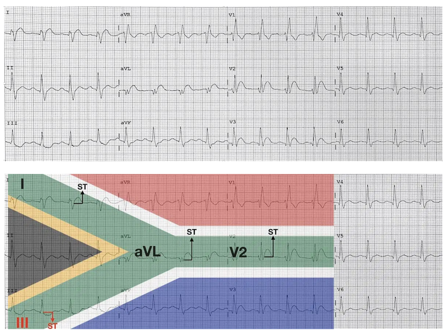
South african flag sign